


Home / Training / Manuals / Atlas of breast cancer early detection / Cases
Atlas of breast cancer early detection
Go back to the list of case studies
.png) Click on the pictures to magnify and display the legends
Click on the pictures to magnify and display the legends
| Case number: | 065 |
| Age: | 60 |
| Clinical presentation: | Postmenopausal woman with average risk of developing breast cancer presented with painless right breast lump. On examination, a hard non-tender lump was found in the right breast. |
Mammography:
 | 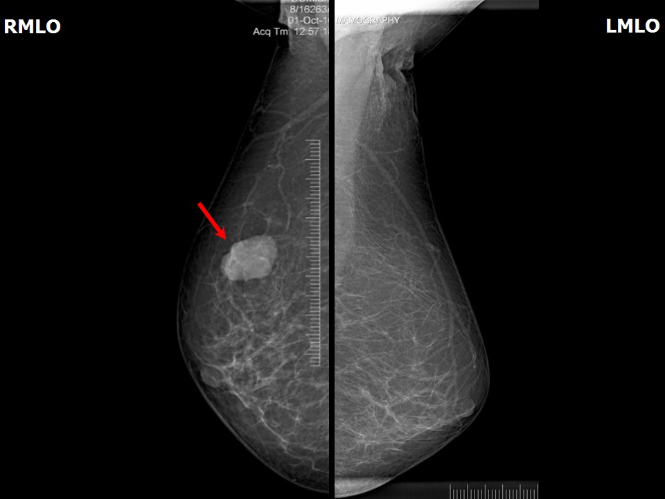 |
| Breast composition: | ACR category a (the breasts are almost entirely fatty) | Mammography features: |
| ‣ Location of the lesion: | Right breast, upper outer quadrant at 10 oclock, middle third |
| ‣ Mass: | |
| • Number: | 1 |
| • Size: | 3.1 × 2.2 cm |
| • Shape: | Oval |
| • Margins: | Partly circumscribed and partly indistinct |
| • Density: | High |
| ‣ Calcifications: | |
| • Typically benign: | None |
| • Suspicious: | None |
| • Distribution: | None |
| ‣ Architectural distortion: | None |
| ‣ Asymmetry: | None |
| ‣ Intramammary node: | None |
| ‣ Skin lesion: | None |
| ‣ Solitary dilated duct: | None |
| ‣ Associated features: | None |
Ultrasound:
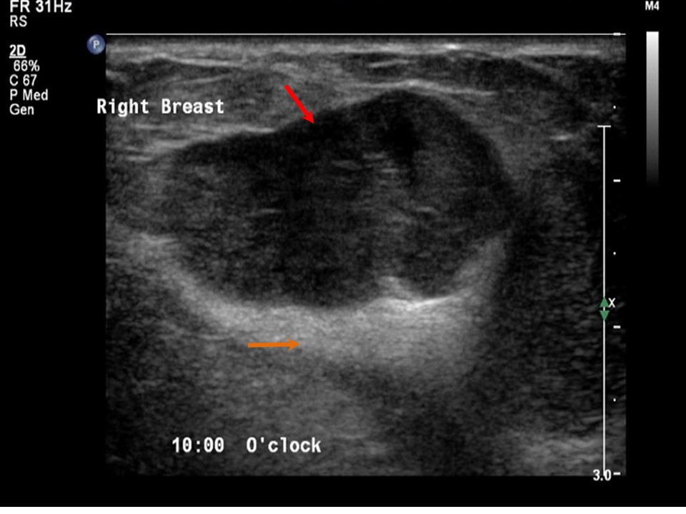 |  |
| Ultrasound features: Right breast, upper outer quadrant at 10 oclock | |
| ‣ Mass | |
| • Location: | Right breast, upper outer quadrant at 10 oclock |
| • Number: | 1 |
| • Size: | 3.0 × 1.4 cm |
| • Shape: | Irregular |
| • Orientation: | Parallel |
| • Margins: | Partly circumscribed and partly indistinct margins |
| • Echo pattern: | Hypoechoic |
| • Posterior features: | Posterior shadowing |
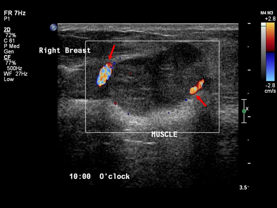
| ‣ Calcifications: | None |
| ‣ Associated features: | Internal vascularity |
| ‣ Special cases: | None |
BI-RADS:
BI-RADS Category: 4B (moderate suspicion of malignancy)Further assessment:
Further assessment advised: Referral for cytologyCytology:
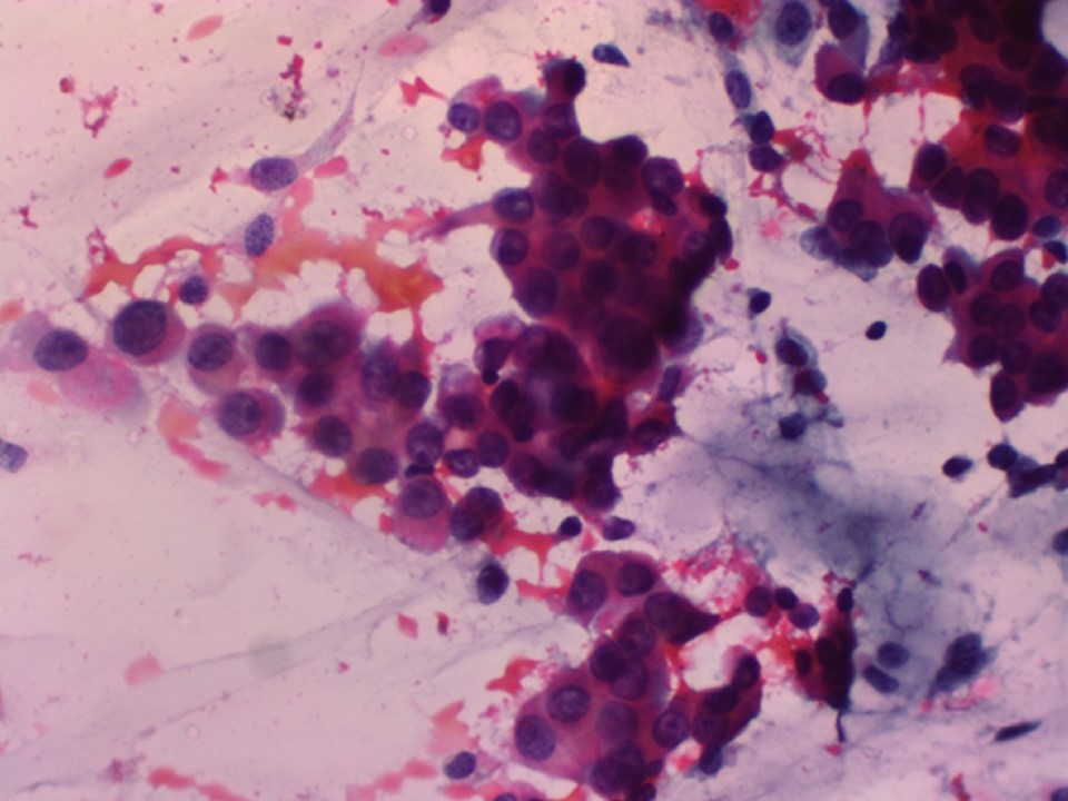 |
| Cytology features: | |
| ‣ Type of sample: | FNAC (solid lesion) |
| ‣ Site of biopsy: | |
| • Laterality: | Right |
| • Quadrant: | |
| • Localization technique: | Palpation |
| • Nature of aspirate: | Mucoid |
| ‣ Cytological description: | Smears show malignant cells with a uniform regular nucleus; many cells are plasmacytoid with eccentric nuclei. Background shows mucinous material |
| ‣ Reporting category: | Malignant |
| ‣ Diagnosis: | Carcinoma |
| ‣ Comments: | None |
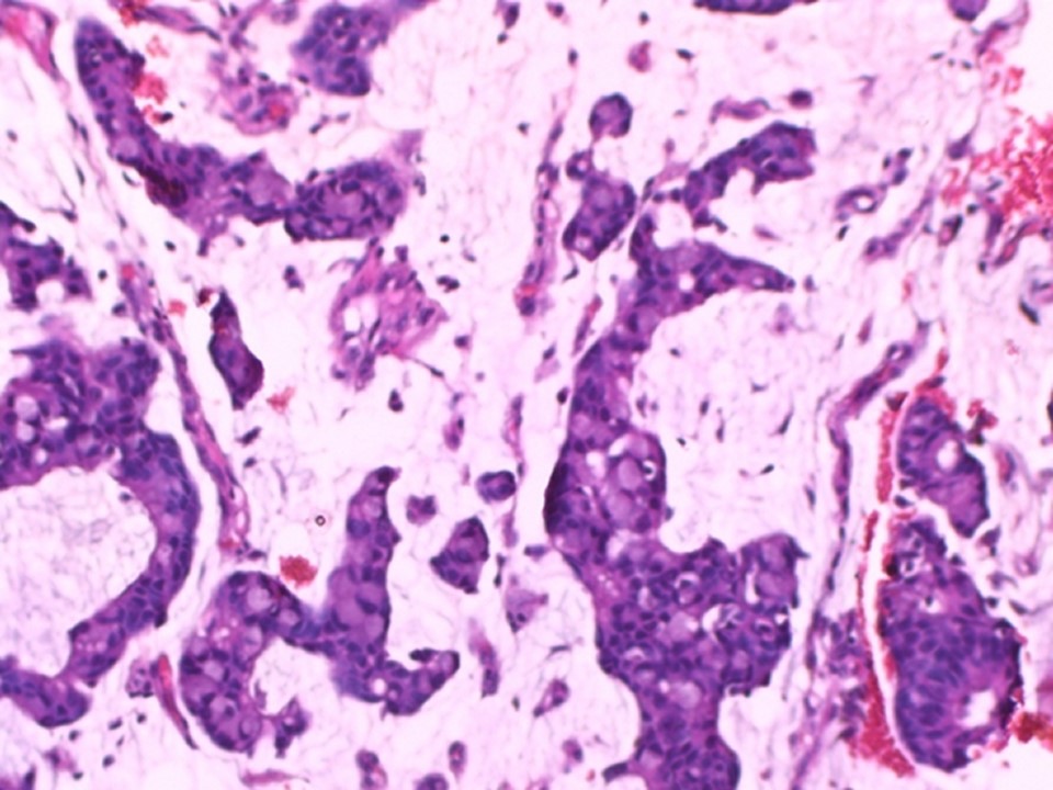
Histopathology:
MRM
 |
| Histopathology features: | |
| ‣ Specimen type: | MRM |
| ‣ Laterality: | Right |
| ‣ Macroscopy: | Cut section shows a well-circumscribed greyish white tumour (2.2 × 2.0 × 1.5 cm), with mucinous areas of soft consistency |
| ‣ Histological type: | Mucinous |
| ‣ Histological grade: | Grade 1 (3 + 1 + 1 = 5) |
| ‣ Mitosis: | Not identified |
| ‣ Maximum invasive tumour size: | 2.2 cm in greatest dimension |
| ‣ Lymph node status: | 0/22 |
| ‣ Peritumoural lymphovascular invasion: | Not identified |
| ‣ DCIS/EIC: | Not identified |
| ‣ Margins: | Free of tumour |
| ‣ Pathological stage: | pT2N0 |
| ‣ Biomarkers: | |
| ‣ Comments: |
Case summary:
| Postmenopausal woman presented with lump in the right breast. Diagnosed as right breast mass of suspicious morphology, BI-RADS 4B on imaging, as mucinous carcinoma on cytology, and as mucinous carcinoma, pT2N0 on histopathology. |
Learning points:
|